Diabetes - Symptoms, Causes, Precaution, Methods and more

  DIABETIES :- Diabetes Mellitus is a chronic metabolic disease. which is characterised by hyperglycemia resulting from insulin deficiency or insulin resistance. Diabetic Mellitus SYMPTOMS :- 3P :-  Polyuria (frequent urine)   Polyphagia ( very hungry)  polydipsia ( very thirsty) feeling tired blurry vision weight loss  delay wound healing HYPERGLYCEMIA High blood sugar/ high sugar level in blood. HYPOGLYCEMIA   Low blood sugar with a glucose value of usually less than 70mg/dl CAUSES To high dose of medication ( Insulin/ antidiabetic medication ) Delayed Meals Exercise Alcohol SYMPTOMS Shakiness Anxiety Sweating Irritability or Confusion Fast heart beat Dizziness Hunger and nausea Headaches Weakness Seizure/Unconsciousness TREATMENT Consume simple carbohydrates like half cup sweetened juice, 3 tablespoons of sugar, honey, chocolates or hard candy. Recheck your blood glucose after 15 minutes. If hyperglycemia continues, repeat the glucose supplements. Once...

Anatomy Of Human Brain, Parts, functions, Lobes, Cranial Nerves, Neuron & Diagram

 HUMAN BRAIN 

Brain is a organ of soft nervous tissue contained in the skull of vertebrates, functioning as the coordinating centre of the body.

PARTS OF BRAIN

Human brain is divided into three major parts on the basis of their functions and placement :- 

1. Fore Brain 

2. Mid Brain 

3. Hind Brain 


Diagram of Human Brain
DIAGRAM OF HUMAN BRAIN

1. FORE BRAIN :-  It is the anterior part of the brain 

It has three parts :- 

  • Thalamus
  • Limbic System
  • Cerebrum

THALAMUS :- it is located above the brain stem and between the cerebral and Mid-Brain.

FUNCTION :- It carries sensory information from the body to cerebrum and the limbic system.

HYPOTHALAMUS :- It lies under the thalamus.

FUNCTION :- It connects the nervous system with the endocrine system via pituitary gland.

LIMBIC SYSTEM :- It is arc shaped structure between thalamus and cerebrum.

FUNCTION :- It controls responses like :-

  • Hunger
  • Fear
  • Thirst
  • Anger 
  • Sexual responses etc.

CEREBRUM :- It is divided into two halves called cerebral hemisphere.
  • They communicate via Corpus Callosum. (Represent white matter)
  • Cerebral Cortex ( Represent gray matter) is the outer region of cerebrum.
FUNCTION :- 
  • It helps in movement
  • It controls speech.
  • It is responsible for sensory processing.
  • It determines the intelligence of the being.

2. MID-BRAIN :- It is located below the cerebral cortex and above the hind brain.
FUNCTION :- It controls reflex movements of the body and hearing reflexes.

3. HIND BRAIN :- It is present at the backside of the brain.
PARTS :- It consist of :- 
  • Cerebellum
  • Pons 
  • Medulla Oblongata

CEREBELLUM :- 
Meaning :- It is latin term for little brain
Position :- It is located at the back side of the head.
function :- It controls the balance of the body and co-ordinates the voluntary movement of the body.

PONS :- 
Meaning :- Pons mean - "Bridge"
Position :- It is located above medulla Oblongata.
Function :- It controls sleep as well as rate and pattern of breathing.

MEDULLA OBLONGATA :- It is the posterior part of the brain.
FUNCTION :- It controls automatic actions example :- Breathing, Heart Rate, Swallowing, Circulation etc 


BRAIN LOBES

Brain is divided into four lobes :- 
  1. Frontal Lobe 
  2.  Parietal Love
  3. Occipital Lobe
  4. Temporal Lobe
Diagram of Brain Lobes
BRAIN LOBES DIAGRAM


CRANIAL NERVES

There are 12 pairs of cranial nerves originating from the nuclei in the inferior surface of brain.
Some are sensory, some are motor and some are mixed.


Cranial nerves
12 Pairs of Cranial Nerves


Cranial nerves Diagram
Diagram of Cranial Nerves


NEURONS

NEURONS :- The neural System of all animals is composed of highly specialised cells called neurons.
FUNCTION :- Which can detect, receive and transmit different kinds of stimuli.

A Neuron is a microscopic structure composed of three major parts :-
1. Cell Body
2. Dendrites 
3. Axon 

CELL BODY :- 
The cell body contains cytoplasm with typical cell organelles and certain granular bodies called Nissl's granules.

DENDRITES :- 
Short branch repeatedly and project out of the cell body and also contain Nissl's granules and are called dendrites.

FUNCTION :- These fibres transmit nerve impulses towards the cell body.

AXON :- 
It is a long fibre, the distal end of which is branched.
FUNCTION :- Axon transmit nerve impulses away from the cell body to a synapse or to a neuro-muscular junction.

SYNAPTIC KNOB :- 
It is a bulb like-structure, possess synaptic vesicles containing chemicals called neurotransmitters.


Structure of Neuron
Diagram of Neuron


NOTE :- Based on the number of axon and dendrites, the Neurons are divided into four types :- 
1. Multipolar
2. Bipolar
3. Unipolar
4. Pseudounipolar 

MULTIPOLAR :- One axon and two or more dendrites, found in the cerebral cortex.

BIPOLAR :- One axon and one dendrites, found in the retina of eye. 

UNIPOLAR :- Cell body with one axon only found usually in embryonic stage.

PSEUDOUNIPOLAR :- It is a sensory Neurons, which contain one axon and split into two parts, which one go into periferal and other one into spinal cord. 

NOTE :- There are two types of Axon 
1. Myelinated
2. Non-Myelinated 

MYELINATED AXON :- Myelinated nerve fibres are enveloped with Schwann cells.
which form a myelin sheath around the axon.
found in spinal and cranial nerves.

NOTE :- The gapes between two adjacent myelin sheaths are called Nodes of Ranvier.

UNMYELINATED AXON :- Unmyelinated nerve fibre is enclosed by a Schwann cells that does not form a myelin sheath around the axon.
found in autonomous and the somatic neural System.


Diagram of Myelinated Axon and Non Myelinated Axon
Structure of Myelinated Axon and Non Myelinated Axon


NEUROGLIA ( NEUROGLIA CELLS )

Glial cells, sometimes called neuroglia or simply glia are non-neuronal cells that maintain homeostasis, from myelin, and protect, support and protection for neurons.

TYPES OF NEUROGLIA CELLS 

There are four types of neuroglia cells :- 
1. Ependymal cells
2. Astrocytes 
3. Microglial cells 
4. Oligodendrocytes

EPENDYMAL CELLS (Light Pink)
Ependymal cells are ciliated-epithelial glial cells that develop from radial glia along the surface of the ventricles of the brain and the spinal canal.

FUNCTION:- Move cerebrous spinal fluid around to keep it homogeneous.
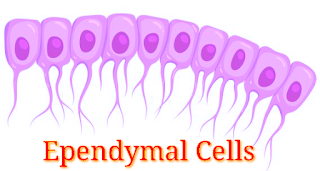
Structure of Ependymal cells


ASTROCYTES (Green)
Astrocytes are specialized glial cells that outnumber neurons by over fivefold.
FUNCTION:- transport of blood-borne material to the neuron, and reaction to injury.
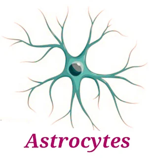
Structure of Astrocytes
Structure of Astrocytes 


MICROGLIAL CELLS (Dark Red)
Microglia cells are the immune cells of the central nervous system.
FUNCTION :- They do phagocytosis to fight infection.

Structure of Microglial cells


OLIGODENDROCYTES (Light Blue)
Oligodendrocytes are the myelinating cells of the central nervous system (CNS).
FUNCTION :- bind the CNS Neurons together and insulate the axons. 

Structure of Oligodendrocytes


Popular posts from this blog

HUMAN CIRCULATORY SYSTEM/BLOOD VASCULAR SYSTEM

HEART CIRCULATORY SYSTEM OR  BLOOD VASCULAR SYSTEM The study of blood vascular system or circulatory system is called angiology    The vascular system is made up of the vessels that carry blood and lymph fluid through the body. It's also called the circulatory system. The arteries and veins carry blood all over the body. They send oxygen and nutrients to the body tissues.   BLOOD VESSELS 1  Arteries  2  Veins 3  Capillaries Differentiate between arteries and vein  CAPILLARIES They have only one layer i.e. simple squamous endothelium layer.  ANATOMY OF HEART Heart  is a coned shaped hollow structure equal to the size of puzzle fit. It is a mesodermally derived organ, is situated in the thoracic cavity, in between the two lungs (mediastinum), slightly tilled to the left. The heart weight between 200 to 425g.  The lower portion is apex and upper portion is base. Wall of heart  1.  Pericardium 2.  Myocardium 3....

ECG Interpretation/ How to read an ECG or EKG/ Characteristics of the normal ECG

  ECG INTERPRETATION BASIC  An ECG complex represents the electrical events occurring in one cardiac cycle. A complex consists of five waveforms labelled with the letters P, Q, R, S, and T. The middle three letters —Q, R, and S—are referred to as a unit, the QRS complex. ECG tracings represent the conduction of electrical impulses from the atria to the ventricles.    P WAVE  Small, Positive and smooth wave. Always +ve in lead II during sinus rhythm. P wave is virtually always +ve in leads aVL, aVF, I, V4, V5, and V6 and -ve in aVR lead. Frequently biphasic in V1 ( occasionally in V2)  the -ve deflection is normally <1mm. P wave duration should be < 0.12 sec. P wave amplitude should be <2.5 mm. Pathological P wave  Tall Tented  :-  Right Atrial Enlargement Bifid (looks like an M) :- Left Atrial Enlargement  PR INTERVAL Normal PR Interval :-  3-5 small squares                  ...

What is Cancer/Types, Symptoms, Causes, Treatment and Diagnosis of Cancer/ What is Silent Cancer

 CANCER  Cancer is also known as Tumor, and study of cancer is called oncology . Definition  Cancer means uncontrol and abnormal division of cells. They are very rapidly proliferate without any differentiation and apoptosis process. (Abnormal proliferative cell is called Neoplastic cell ) Or The uncontrolled growth of cell by its multiplication is known as Cancer. Types of cancer  There are two types of Tumor :- 1. Benign Tumor  2. Malignant Tumor Benign Tumor  Benign Tumor is also called mass of cell. It cannot spread from one location to other location. That means it is not show metastasis. This is a localised tumor. That means it is present only one location not spread to other location. Benign Tumor (Cancer) This type of tumor can remove from the body because it is located only on surface of organ. Example of Benign Tumor :-  Lipoma Myloma  Malignant Tumor  Malignant Tumor is very dangerous tumor because it can spread or can transfer from...

ELECTROCARDIOGRAPHY BASIC

  What is electrocardiography (ECG) Electrocardiography is a medical test in which diagnose cardiac disorder i.e. MI (myocardial infarction) etc. by measuring the electrical activity of the heart via electrodes placed on the limbs and chest wall and are transcribed on to graph paper to produce an electrocardiogram and also known as ECG/EKG.                                     OR     Electrocardiogram   Electro means electrical activity   Cardio means heart   Gram means graphical representation   An electrocardiogram is a graphically representation of an electrical activity of the heart and used in the investigation of heart disease.   The device is used is known as electrocardiograph.   NOTE -- The term electrocardiogram was introduced by Willem Einthoven ...

Atrial Fibrillation/Atrial Flutter

  Atrial Fibrillation  Atrial Flutter Atrial Fibrillation   (Afib) Afib stands for atrial Fibrillation (AF) Atrial Fibrillation is a type of arrhythmia or abnormal heart beat and is a major cause of stroke.  Atrial Fibrillation is an irregularly irregular and often very rapid heart rhythm that begins in heart upper chambers (Atria). in which the upper chambers of the heart (atria) beat out of coordination with the lower chambers of the heart (ventricle). ATRIAL FIBRILLATION FINDINGS ON ECG Findings Irregularly irregular QRS Complex P wave absent / no visible P wave present Heart beat range (100bpm - 160bpm) Types of Atrial Fibrillation There are three main types of atrial fibrillation.... Paroxysmal atrial Fibrillation Persistent atrial fibrillation Long standing persistent atrial fibrillation Paroxysmal Atrial Fibrillation It's episodes occurs less than one week and usually stops on its own without treatment. Persistent Atrial Fibrillation It's episodes occurs more ...