Showing posts with label Anatomy of heart. Show all posts

What is Cancer/Types, Symptoms, Causes, Treatment and Diagnosis of Cancer/ What is Silent Cancer

 CANCER 

Cancer is also known as Tumor, and study of cancer is called oncology.

Definition 

Cancer means uncontrol and abnormal division of cells. They are very rapidly proliferate without any differentiation and apoptosis process.
(Abnormal proliferative cell is called Neoplastic cell)

Or

The uncontrolled growth of cell by its multiplication is known as Cancer.


Types of cancer 

There are two types of Tumor :-

1. Benign Tumor 
2. Malignant Tumor

Benign Tumor 

Benign Tumor is also called mass of cell.
It cannot spread from one location to other location.
That means it is not show metastasis.

This is a localised tumor. That means it is present only one location not spread to other location.

Benign Tumor (Cancer)

Benign Tumor (Cancer)


This type of tumor can remove from the body because it is located only on surface of
organ.


Example of Benign Tumor :- 

Lipoma
Myloma 

Malignant Tumor 

Malignant Tumor is very dangerous tumor because it can spread or can transfer from one location to other location.

Malignant Tumor show metastasis process.

Malignant Tumor (cancer cells)
Malignant Tumor (Cancer)


Types of Malignant Tumor :- 

There are four types of Malignant Tumor/Cancer :-

1. Carcinoma 

It is also known as skin cancer occur in epithelial tissue, cause by ultraviolet rays of sun.
Carcinoma (Malignant Tumor)

Carcinoma (Malignant Tumor)
Carcinoma 


2. Sarcoma 

Cancerous growth in connective tissue, bones, cartilage and muscle is called sarcoma.
Sarcoma (Cancer) Sarcoma

Sarcoma (Cancer)
Sarcoma 


3. Leukemia 

Abnormal growth in the number of leucocytes, also known as blood cancer.
Leukemia (Blood cancer)

Leukemia (Blood cancer)

Leukemia (Blood Cancer)

Leukemia (Blood Cancer)


4. Lymphoma 

Cancerous growth occur in lymph node or spleen.

Lymphoma (Cancer)

Lymphoma (Cancer)
Lymphoma 


Symptoms of Cancer :-

  • Fatigue 
  • Unintentional weight loss
  • Unexplained fever 
  • Changes in bowel 
  • Abnormal lumps
  • Persistent cough
  • Unexplained muscles and joint pain 
  • Night Sweat
  • Digestion problem 
  • Discomfort after eating 
  • Skin color changes occur - Redness of the skin
  • Nails color changes occur 
  • Unusual bleeding 
  • Bowel habit change 

Causes of Cancer 

A special agent which can cause a cancer that is called Carcinogen
  • Physical agent 
  • Chemical agent
  • Biological agent
  • Radiation 

Physical and Chemical agent 

  • Tobacco 
  • Smoking 
  • Alcohol 

Biological agent 

  • Parasite 
Schistosoma parasite cause cancer.

  • Virus 
Hepatitis B and C virus can cause cancer.
Virus which can cause a cancer is called oncogenic virus.

Radiation 

X-Rays
Gama Rays
UV Rays


What is silent cancer ?

Silent cancer do not have initial symptoms, 
This is because, most of these cancer affect internal organs and deep inside the body such as lungs, intestine, liver, bladder and pancreas etc. 
That's why it is difficult to find them during a regular physical exercise.


Treatment of cancer 

1. Surgery 

Benign Tumor Cancer easy remove by surgery.

2. Radiation Therapy 

Different types of radiation are used for the treatment of cancer.
In a radiation Therapy radiation stops the growth of cancer cells without any harms.

3. Chemotherapy 

It is a process in which different Antinuclear drugs are used which is inhibit a cancer cell DNA.

Chemotherapy has some minor disadvantages:- 
Hair loss
Weakness 
Anemia 

How can check you have cancer or not ?

Common test to diagnose cancer :-

  • Blood Chemistry Test
  • CBC (Complete blood count)
  • Genetic test
  • Urinalysis 
  • CT Scan
  • MRI
  • Ultrasound 
  • X-Ray
  • Biopsy 
  • Tumor marker test


HUMAN CIRCULATORY SYSTEM/BLOOD VASCULAR SYSTEM

HEART CIRCULATORY SYSTEM

OR 

BLOOD VASCULAR SYSTEM


The study of blood vascular system or circulatory system is called angiology  


The vascular system is made up of the vessels that carry blood and lymph fluid through the body. It's also called the circulatory system. The arteries and veins carry blood all over the body. They send oxygen and nutrients to the body tissues.  

Blood Vascular System

BLOOD VESSELS

Arteries 

Veins

Capillaries

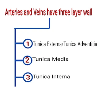
All Layer of Arteries and Veins
Diagram of T.S. of Arteries and T.S. of Veins


Differentiate between arteries and vein 


Difference between Arteries and Veins


CAPILLARIES

They have only one layer i.e. simple squamous endothelium layer. 

Diagram of T.S. of Capillaries


ANATOMY OF HEART


Heart  is a coned shaped hollow structure equal to the size of puzzle fit.

It is a mesodermally derived organ, is situated in the thoracic cavity, in between the two lungs (mediastinum), slightly tilled to the left.

The heart weight between 200 to 425g. 
The lower portion is apex and upper portion is base.

Wall of heart 

1.  Pericardium

2.  Myocardium

3.  Endocardium

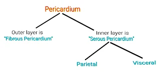
Pericardium-- It is a outermost layer of the heart.
Heart is protected by a double walled membranous bag like structure called pericardium.

Pericardium layer of Heart

Myocardium -- Consist of specialized cardiac muscle (involuntary in nature) found only in the heart.

Endocardium -- It consist of flatted epithelial cell and it is continuous with endothelium lining the blood vessels.

Labelled Diagram of Heart


Mammals heart have a four chambers, two small upper chambers called atria and two large lower chambers called ventricles.

Inter-atrial septum wall separate the right and the left atria.
It is a thin muscular layer (wall).

Inter-ventricular septum wall separate the right and left ventricles.
It is a thick muscular wall.

Atrio-ventricualr septum separate the atrium and ventricles of the same side.

Blood Circulation of Heart


AORTIC ARCH


Aortic Arch
The aorta arch is the section of the aorta between the ascending and descending aorta that distribute blood to the head and upper extremities via the brachiocephalic trunk, the left common carotid artery, and the left subclavian artery. 

The aortic arch has 3 major branches.
The brachiocephalic trunk is the first branch of the aortic arch and supplies blood to the right arm and right head and neck.
The left common carotid artery is the second branch of the aortic arch, which supplies blood to the left side of the head and the neck.
The last branch of the aortic arch is the left subclavian artery that distribute blood to the left arm. 


PULMONARY CIRCULATION


The deoxygenated blood pumped into the pulmonary artery is passed on to the lungs (from where the oxygenated blood is carried by the pulmonary veins into the left atrium) this pathway is called pulmonary circulation.

Pulmonary circulation occur only between the heart and lungs. 


SYSTEMIC CIRCULATION


Circulation of blood throughout the body through the arteries, capillaries and veins, which carry oxygenated blood from the left ventricle to various tissue and return venous blood to the right atrium.

Systemic circulation occur between the heart and body. 

Diagram of pulmonary circulation and systemic circulation


CONDUCTION SYSTEM OF HEART


The cardiac conduction system is a group of specialized cardiac muscle cells in the walls of the heart.
That send signals to the heart muscle causing it to contract.

The main components of the cardiac conduction system are the SA Node, AV Node, bundle of his, bundle branches and Purkinje fibers.

Conduction system of heart


Diagram of Conduction system of heart

Diagram of Conduction system of heart


FUNCTION OF SA NODE

(Sino-atrial node) or (Natural pacemaker of heart)


1. The SAN can generate the maximum number of action potential. i.e. 70-75 beats per minute (bpm).

2. It is responsible for initiating and maintaining the rhythmic contractile activity of the heart that's why it is called the natural pacemaker.

NOTE- Our heart normally beats 70-75 times in a minute.
(Average 72 beats per minute)


FUNCTION OF AV NODE


1. The purpose of this structure is to connect the electrical system of the atria and the Ventricles, providing electrical impedance from the atria and an intrinsic pacemaker in its absence.

2. The intrinsic rate of the AV node is 40-60 beats per minute (bpm).


Diabetes - Symptoms, Causes, Precaution, Methods and more

  DIABETIES :- Diabetes Mellitus is a chronic metabolic disease. which is characterised by hyperglycemia resulting from insulin deficiency ...