Understanding Cancer: Key Facts and Prevention Tips for Everyone


Spreading Hope & Awareness: Understanding Cancer Better


Cancer. A word that stirs deep emotions—fear, sadness, and often a fight for life. Yet, it's a battle that millions have faced and many have survived. This Cancer Awareness post is dedicated to spreading knowledge and hope, emphasizing the importance of early detection, lifestyle choices, and support systems in fighting this disease.


TOGETHER AGAINST CANCER AWARENESS AND HOPE
TOGETHER AGAINST CANCER AWARENESS AND HOPE 


Understanding Cancer:


Pink Minimal Breast Cancer Awareness Campaign Instagram Post by Sonam Saxena

Cancer is not just one disease but a group of over 100 different diseases. It happens when cells in the body grow uncontrollably. These abnormal cells can invade other parts of the body, posing severe health risks. The most common types include breast cancer, lung cancer, skin cancer, and prostate cancer, but it can affect nearly any part of the body.


Why Awareness Matters:


Early detection can be life-saving. Many types of cancer can be treated more effectively if caught early. However, lack of awareness and stigma often prevent people from seeking timely help. Cancer awareness campaigns help educate people about symptoms, risk factors, and screening methods that could save lives.


Prevention and Early Detection:


Regular Screenings: Regular health check-ups, including mammograms, colonoscopies, and skin checks, can detect cancer at an early in a balanced diet, avoiding smoking, reducing alcohol intake, and staying physically active—can reduce the risk of many cancers.


Know the Symptoms: Unusual lumps, unexplained weight loss, persistent cough, or changes in moles are signs that shouldn't be ignored. Consult a doctor if you notice these.


Common Cancer Symptoms: Recognizing Hidden Illness

Cancer is a disease that often doesn’t show specific symptoms in its early stages, making awareness and timely recognition crucial. In this post, we’ll explore some common cancer symptoms in detail, helping you understand them better and encouraging you to consult a doctor if necessary.


1. Sudden Weight Loss

If you experience unexpected weight loss without any changes to your diet or exercise routine, this could be a concern. This symptom is often associated with cancer, particularly gastrointestinal cancers (like stomach cancer). Rapid growth of cancer cells can outpace the body's normal cells, leading to unexplained weight loss.


What to Do? If you notice a 5-10% weight loss over a few months without any intentional efforts, it’s essential to pay attention to this symptom. Consider consulting a doctor for further evaluation.


2. Persistent Fatigue

Constant fatigue is another common symptom of cancer. This type of fatigue is not relieved by rest and can significantly affect your daily life. Cancer-related fatigue is often due to cancer cells consuming energy and nutrients from the body.


What to Do? If this fatigue is impacting your daily activities and persists over time, don’t hesitate to reach out to a healthcare professional.


3. Persistent Cough or Blood in Cough

A persistent cough or coughing up blood can be serious and is often associated with certain types of cancers, like lung cancer. This symptom should not be overlooked as it can indicate underlying health issues.


What to Do? If you have a cough that lasts more than a month or you notice blood in your sputum, it’s crucial to seek medical attention immediately.


4. Loss of Appetite or Unexplained Changes in Eating Habits

A sudden lack of interest in food or significant changes in appetite can be warning signs of cancer. This can happen due to hormonal changes or the body’s response to cancer.


What to Do? If you experience a marked change in your appetite or find yourself eating much less without a clear reason, consult a healthcare provider for advice.


5. Unexplained Lumps or Swelling

Finding a lump or experiencing unusual swelling in any part of your body can be a warning sign of cancer. This is especially true for cancers like lymphoma or breast cancer, where swelling may occur in the lymph nodes or breast tissue.


What to Do? If a lump or swelling persists for more than a month or continues to grow, it’s important to see a doctor for evaluation. They can perform tests to determine the cause.


Conclusion

Understanding cancer symptoms and recognizing them early is vital for timely diagnosis and treatment. If you experience any of these symptoms, don’t hesitate to contact a healthcare professional. Awareness and early intervention can significantly impact health outcomes. Taking care of your health and the health of your loved ones should always be a priority.


Supporting Those in the Fight:


Cancer can be a lonely journey, but nobody should face it alone. Family, friends, and even online support groups play a vital role in providing emotional and psychological support. As a society, let’s show compassion, reduce stigma, and help patients feel they are not alone in their fight.


How Can You Help?


Donate to Cancer Research: Funds are critical for advancing treatments and finding cures.


Volunteer Time: Even small acts of kindness can make a big difference for patients and their families.


Spread the Word: Share posts, articles, and reliable information about cancer on your social media to educate more people.


Together, We Are Stronger


Cancer is a formidable adversary, but the human spirit is stronger. By staying informed, spreading awareness, and supporting each other, we can face this challenge together. Let’s make every day a step towards a cancer-free world.


Anatomy Of Human Brain, Parts, functions, Lobes, Cranial Nerves, Neuron & Diagram

 HUMAN BRAIN 

Brain is a organ of soft nervous tissue contained in the skull of vertebrates, functioning as the coordinating centre of the body.

PARTS OF BRAIN

Human brain is divided into three major parts on the basis of their functions and placement :- 

1. Fore Brain 

2. Mid Brain 

3. Hind Brain 


Diagram of Human Brain
DIAGRAM OF HUMAN BRAIN

1. FORE BRAIN :-  It is the anterior part of the brain 

It has three parts :- 

  • Thalamus
  • Limbic System
  • Cerebrum

THALAMUS :- it is located above the brain stem and between the cerebral and Mid-Brain.

FUNCTION :- It carries sensory information from the body to cerebrum and the limbic system.

HYPOTHALAMUS :- It lies under the thalamus.

FUNCTION :- It connects the nervous system with the endocrine system via pituitary gland.

LIMBIC SYSTEM :- It is arc shaped structure between thalamus and cerebrum.

FUNCTION :- It controls responses like :-

  • Hunger
  • Fear
  • Thirst
  • Anger 
  • Sexual responses etc.

CEREBRUM :- It is divided into two halves called cerebral hemisphere.
  • They communicate via Corpus Callosum. (Represent white matter)
  • Cerebral Cortex ( Represent gray matter) is the outer region of cerebrum.
FUNCTION :- 
  • It helps in movement
  • It controls speech.
  • It is responsible for sensory processing.
  • It determines the intelligence of the being.

2. MID-BRAIN :- It is located below the cerebral cortex and above the hind brain.
FUNCTION :- It controls reflex movements of the body and hearing reflexes.

3. HIND BRAIN :- It is present at the backside of the brain.
PARTS :- It consist of :- 
  • Cerebellum
  • Pons 
  • Medulla Oblongata

CEREBELLUM :- 
Meaning :- It is latin term for little brain
Position :- It is located at the back side of the head.
function :- It controls the balance of the body and co-ordinates the voluntary movement of the body.

PONS :- 
Meaning :- Pons mean - "Bridge"
Position :- It is located above medulla Oblongata.
Function :- It controls sleep as well as rate and pattern of breathing.

MEDULLA OBLONGATA :- It is the posterior part of the brain.
FUNCTION :- It controls automatic actions example :- Breathing, Heart Rate, Swallowing, Circulation etc 


BRAIN LOBES

Brain is divided into four lobes :- 
  1. Frontal Lobe 
  2.  Parietal Love
  3. Occipital Lobe
  4. Temporal Lobe
Diagram of Brain Lobes
BRAIN LOBES DIAGRAM


CRANIAL NERVES

There are 12 pairs of cranial nerves originating from the nuclei in the inferior surface of brain.
Some are sensory, some are motor and some are mixed.


Cranial nerves
12 Pairs of Cranial Nerves


Cranial nerves Diagram
Diagram of Cranial Nerves


NEURONS

NEURONS :- The neural System of all animals is composed of highly specialised cells called neurons.
FUNCTION :- Which can detect, receive and transmit different kinds of stimuli.

A Neuron is a microscopic structure composed of three major parts :-
1. Cell Body
2. Dendrites 
3. Axon 

CELL BODY :- 
The cell body contains cytoplasm with typical cell organelles and certain granular bodies called Nissl's granules.

DENDRITES :- 
Short branch repeatedly and project out of the cell body and also contain Nissl's granules and are called dendrites.

FUNCTION :- These fibres transmit nerve impulses towards the cell body.

AXON :- 
It is a long fibre, the distal end of which is branched.
FUNCTION :- Axon transmit nerve impulses away from the cell body to a synapse or to a neuro-muscular junction.

SYNAPTIC KNOB :- 
It is a bulb like-structure, possess synaptic vesicles containing chemicals called neurotransmitters.


Structure of Neuron
Diagram of Neuron


NOTE :- Based on the number of axon and dendrites, the Neurons are divided into four types :- 
1. Multipolar
2. Bipolar
3. Unipolar
4. Pseudounipolar 

MULTIPOLAR :- One axon and two or more dendrites, found in the cerebral cortex.

BIPOLAR :- One axon and one dendrites, found in the retina of eye. 

UNIPOLAR :- Cell body with one axon only found usually in embryonic stage.

PSEUDOUNIPOLAR :- It is a sensory Neurons, which contain one axon and split into two parts, which one go into periferal and other one into spinal cord. 

NOTE :- There are two types of Axon 
1. Myelinated
2. Non-Myelinated 

MYELINATED AXON :- Myelinated nerve fibres are enveloped with Schwann cells.
which form a myelin sheath around the axon.
found in spinal and cranial nerves.

NOTE :- The gapes between two adjacent myelin sheaths are called Nodes of Ranvier.

UNMYELINATED AXON :- Unmyelinated nerve fibre is enclosed by a Schwann cells that does not form a myelin sheath around the axon.
found in autonomous and the somatic neural System.


Diagram of Myelinated Axon and Non Myelinated Axon
Structure of Myelinated Axon and Non Myelinated Axon


NEUROGLIA ( NEUROGLIA CELLS )

Glial cells, sometimes called neuroglia or simply glia are non-neuronal cells that maintain homeostasis, from myelin, and protect, support and protection for neurons.

TYPES OF NEUROGLIA CELLS 

There are four types of neuroglia cells :- 
1. Ependymal cells
2. Astrocytes 
3. Microglial cells 
4. Oligodendrocytes

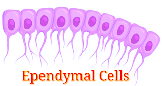
EPENDYMAL CELLS (Light Pink)
Ependymal cells are ciliated-epithelial glial cells that develop from radial glia along the surface of the ventricles of the brain and the spinal canal.

FUNCTION:- Move cerebrous spinal fluid around to keep it homogeneous.

Structure of Ependymal cells


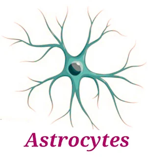
ASTROCYTES (Green)
Astrocytes are specialized glial cells that outnumber neurons by over fivefold.
FUNCTION:- transport of blood-borne material to the neuron, and reaction to injury.

Structure of Astrocytes
Structure of Astrocytes 


MICROGLIAL CELLS (Dark Red)
Microglia cells are the immune cells of the central nervous system.
FUNCTION :- They do phagocytosis to fight infection.

Structure of Microglial cells


OLIGODENDROCYTES (Light Blue)
Oligodendrocytes are the myelinating cells of the central nervous system (CNS).
FUNCTION :- bind the CNS Neurons together and insulate the axons. 

Structure of Oligodendrocytes


Understanding Cancer: Key Facts and Prevention Tips for Everyone

  Spreading Hope & Awareness: Understanding Cancer Better Cancer . A word that stirs deep emotions—fear, sadness, and often a fight for ...