Understanding Cancer: Key Facts and Prevention Tips for Everyone


Spreading Hope & Awareness: Understanding Cancer Better


Cancer. A word that stirs deep emotions—fear, sadness, and often a fight for life. Yet, it's a battle that millions have faced and many have survived. This Cancer Awareness post is dedicated to spreading knowledge and hope, emphasizing the importance of early detection, lifestyle choices, and support systems in fighting this disease.


TOGETHER AGAINST CANCER AWARENESS AND HOPE
TOGETHER AGAINST CANCER AWARENESS AND HOPE 


Understanding Cancer:


Pink Minimal Breast Cancer Awareness Campaign Instagram Post by Sonam Saxena

Cancer is not just one disease but a group of over 100 different diseases. It happens when cells in the body grow uncontrollably. These abnormal cells can invade other parts of the body, posing severe health risks. The most common types include breast cancer, lung cancer, skin cancer, and prostate cancer, but it can affect nearly any part of the body.


Why Awareness Matters:


Early detection can be life-saving. Many types of cancer can be treated more effectively if caught early. However, lack of awareness and stigma often prevent people from seeking timely help. Cancer awareness campaigns help educate people about symptoms, risk factors, and screening methods that could save lives.


Prevention and Early Detection:


Regular Screenings: Regular health check-ups, including mammograms, colonoscopies, and skin checks, can detect cancer at an early in a balanced diet, avoiding smoking, reducing alcohol intake, and staying physically active—can reduce the risk of many cancers.


Know the Symptoms: Unusual lumps, unexplained weight loss, persistent cough, or changes in moles are signs that shouldn't be ignored. Consult a doctor if you notice these.


Common Cancer Symptoms: Recognizing Hidden Illness

Cancer is a disease that often doesn’t show specific symptoms in its early stages, making awareness and timely recognition crucial. In this post, we’ll explore some common cancer symptoms in detail, helping you understand them better and encouraging you to consult a doctor if necessary.


1. Sudden Weight Loss

If you experience unexpected weight loss without any changes to your diet or exercise routine, this could be a concern. This symptom is often associated with cancer, particularly gastrointestinal cancers (like stomach cancer). Rapid growth of cancer cells can outpace the body's normal cells, leading to unexplained weight loss.


What to Do? If you notice a 5-10% weight loss over a few months without any intentional efforts, it’s essential to pay attention to this symptom. Consider consulting a doctor for further evaluation.


2. Persistent Fatigue

Constant fatigue is another common symptom of cancer. This type of fatigue is not relieved by rest and can significantly affect your daily life. Cancer-related fatigue is often due to cancer cells consuming energy and nutrients from the body.


What to Do? If this fatigue is impacting your daily activities and persists over time, don’t hesitate to reach out to a healthcare professional.


3. Persistent Cough or Blood in Cough

A persistent cough or coughing up blood can be serious and is often associated with certain types of cancers, like lung cancer. This symptom should not be overlooked as it can indicate underlying health issues.


What to Do? If you have a cough that lasts more than a month or you notice blood in your sputum, it’s crucial to seek medical attention immediately.


4. Loss of Appetite or Unexplained Changes in Eating Habits

A sudden lack of interest in food or significant changes in appetite can be warning signs of cancer. This can happen due to hormonal changes or the body’s response to cancer.


What to Do? If you experience a marked change in your appetite or find yourself eating much less without a clear reason, consult a healthcare provider for advice.


5. Unexplained Lumps or Swelling

Finding a lump or experiencing unusual swelling in any part of your body can be a warning sign of cancer. This is especially true for cancers like lymphoma or breast cancer, where swelling may occur in the lymph nodes or breast tissue.


What to Do? If a lump or swelling persists for more than a month or continues to grow, it’s important to see a doctor for evaluation. They can perform tests to determine the cause.


Conclusion

Understanding cancer symptoms and recognizing them early is vital for timely diagnosis and treatment. If you experience any of these symptoms, don’t hesitate to contact a healthcare professional. Awareness and early intervention can significantly impact health outcomes. Taking care of your health and the health of your loved ones should always be a priority.


Supporting Those in the Fight:


Cancer can be a lonely journey, but nobody should face it alone. Family, friends, and even online support groups play a vital role in providing emotional and psychological support. As a society, let’s show compassion, reduce stigma, and help patients feel they are not alone in their fight.


How Can You Help?


Donate to Cancer Research: Funds are critical for advancing treatments and finding cures.


Volunteer Time: Even small acts of kindness can make a big difference for patients and their families.


Spread the Word: Share posts, articles, and reliable information about cancer on your social media to educate more people.


Together, We Are Stronger


Cancer is a formidable adversary, but the human spirit is stronger. By staying informed, spreading awareness, and supporting each other, we can face this challenge together. Let’s make every day a step towards a cancer-free world.


Anatomy Of Human Brain, Parts, functions, Lobes, Cranial Nerves, Neuron & Diagram

 HUMAN BRAIN 

Brain is a organ of soft nervous tissue contained in the skull of vertebrates, functioning as the coordinating centre of the body.

PARTS OF BRAIN

Human brain is divided into three major parts on the basis of their functions and placement :- 

1. Fore Brain 

2. Mid Brain 

3. Hind Brain 


Diagram of Human Brain
DIAGRAM OF HUMAN BRAIN

1. FORE BRAIN :-  It is the anterior part of the brain 

It has three parts :- 

  • Thalamus
  • Limbic System
  • Cerebrum

THALAMUS :- it is located above the brain stem and between the cerebral and Mid-Brain.

FUNCTION :- It carries sensory information from the body to cerebrum and the limbic system.

HYPOTHALAMUS :- It lies under the thalamus.

FUNCTION :- It connects the nervous system with the endocrine system via pituitary gland.

LIMBIC SYSTEM :- It is arc shaped structure between thalamus and cerebrum.

FUNCTION :- It controls responses like :-

  • Hunger
  • Fear
  • Thirst
  • Anger 
  • Sexual responses etc.

CEREBRUM :- It is divided into two halves called cerebral hemisphere.
  • They communicate via Corpus Callosum. (Represent white matter)
  • Cerebral Cortex ( Represent gray matter) is the outer region of cerebrum.
FUNCTION :- 
  • It helps in movement
  • It controls speech.
  • It is responsible for sensory processing.
  • It determines the intelligence of the being.

2. MID-BRAIN :- It is located below the cerebral cortex and above the hind brain.
FUNCTION :- It controls reflex movements of the body and hearing reflexes.

3. HIND BRAIN :- It is present at the backside of the brain.
PARTS :- It consist of :- 
  • Cerebellum
  • Pons 
  • Medulla Oblongata

CEREBELLUM :- 
Meaning :- It is latin term for little brain
Position :- It is located at the back side of the head.
function :- It controls the balance of the body and co-ordinates the voluntary movement of the body.

PONS :- 
Meaning :- Pons mean - "Bridge"
Position :- It is located above medulla Oblongata.
Function :- It controls sleep as well as rate and pattern of breathing.

MEDULLA OBLONGATA :- It is the posterior part of the brain.
FUNCTION :- It controls automatic actions example :- Breathing, Heart Rate, Swallowing, Circulation etc 


BRAIN LOBES

Brain is divided into four lobes :- 
  1. Frontal Lobe 
  2.  Parietal Love
  3. Occipital Lobe
  4. Temporal Lobe
Diagram of Brain Lobes
BRAIN LOBES DIAGRAM


CRANIAL NERVES

There are 12 pairs of cranial nerves originating from the nuclei in the inferior surface of brain.
Some are sensory, some are motor and some are mixed.


Cranial nerves
12 Pairs of Cranial Nerves


Cranial nerves Diagram
Diagram of Cranial Nerves


NEURONS

NEURONS :- The neural System of all animals is composed of highly specialised cells called neurons.
FUNCTION :- Which can detect, receive and transmit different kinds of stimuli.

A Neuron is a microscopic structure composed of three major parts :-
1. Cell Body
2. Dendrites 
3. Axon 

CELL BODY :- 
The cell body contains cytoplasm with typical cell organelles and certain granular bodies called Nissl's granules.

DENDRITES :- 
Short branch repeatedly and project out of the cell body and also contain Nissl's granules and are called dendrites.

FUNCTION :- These fibres transmit nerve impulses towards the cell body.

AXON :- 
It is a long fibre, the distal end of which is branched.
FUNCTION :- Axon transmit nerve impulses away from the cell body to a synapse or to a neuro-muscular junction.

SYNAPTIC KNOB :- 
It is a bulb like-structure, possess synaptic vesicles containing chemicals called neurotransmitters.


Structure of Neuron
Diagram of Neuron


NOTE :- Based on the number of axon and dendrites, the Neurons are divided into four types :- 
1. Multipolar
2. Bipolar
3. Unipolar
4. Pseudounipolar 

MULTIPOLAR :- One axon and two or more dendrites, found in the cerebral cortex.

BIPOLAR :- One axon and one dendrites, found in the retina of eye. 

UNIPOLAR :- Cell body with one axon only found usually in embryonic stage.

PSEUDOUNIPOLAR :- It is a sensory Neurons, which contain one axon and split into two parts, which one go into periferal and other one into spinal cord. 

NOTE :- There are two types of Axon 
1. Myelinated
2. Non-Myelinated 

MYELINATED AXON :- Myelinated nerve fibres are enveloped with Schwann cells.
which form a myelin sheath around the axon.
found in spinal and cranial nerves.

NOTE :- The gapes between two adjacent myelin sheaths are called Nodes of Ranvier.

UNMYELINATED AXON :- Unmyelinated nerve fibre is enclosed by a Schwann cells that does not form a myelin sheath around the axon.
found in autonomous and the somatic neural System.


Diagram of Myelinated Axon and Non Myelinated Axon
Structure of Myelinated Axon and Non Myelinated Axon


NEUROGLIA ( NEUROGLIA CELLS )

Glial cells, sometimes called neuroglia or simply glia are non-neuronal cells that maintain homeostasis, from myelin, and protect, support and protection for neurons.

TYPES OF NEUROGLIA CELLS 

There are four types of neuroglia cells :- 
1. Ependymal cells
2. Astrocytes 
3. Microglial cells 
4. Oligodendrocytes

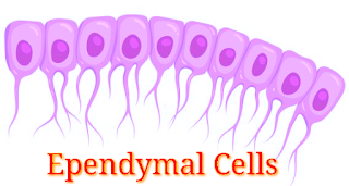
EPENDYMAL CELLS (Light Pink)
Ependymal cells are ciliated-epithelial glial cells that develop from radial glia along the surface of the ventricles of the brain and the spinal canal.

FUNCTION:- Move cerebrous spinal fluid around to keep it homogeneous.

Structure of Ependymal cells


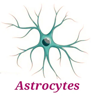
ASTROCYTES (Green)
Astrocytes are specialized glial cells that outnumber neurons by over fivefold.
FUNCTION:- transport of blood-borne material to the neuron, and reaction to injury.

Structure of Astrocytes
Structure of Astrocytes 


MICROGLIAL CELLS (Dark Red)
Microglia cells are the immune cells of the central nervous system.
FUNCTION :- They do phagocytosis to fight infection.

Structure of Microglial cells


OLIGODENDROCYTES (Light Blue)
Oligodendrocytes are the myelinating cells of the central nervous system (CNS).
FUNCTION :- bind the CNS Neurons together and insulate the axons. 

Structure of Oligodendrocytes


What is Cancer/Types, Symptoms, Causes, Treatment and Diagnosis of Cancer/ What is Silent Cancer

 CANCER 

Cancer is also known as Tumor, and study of cancer is called oncology.

Definition 

Cancer means uncontrol and abnormal division of cells. They are very rapidly proliferate without any differentiation and apoptosis process.
(Abnormal proliferative cell is called Neoplastic cell)

Or

The uncontrolled growth of cell by its multiplication is known as Cancer.


Types of cancer 

There are two types of Tumor :-

1. Benign Tumor 
2. Malignant Tumor

Benign Tumor 

Benign Tumor is also called mass of cell.
It cannot spread from one location to other location.
That means it is not show metastasis.

This is a localised tumor. That means it is present only one location not spread to other location.

Benign Tumor (Cancer)
Benign Tumor 


This type of tumor can remove from the body because it is located only on surface of
organ.

Benign Tumor (Cancer)
Benign Tumor 


Example of Benign Tumor :- 

Lipoma
Myloma 

Malignant Tumor 

Malignant Tumor is very dangerous tumor because it can spread or can transfer from one location to other location.

Malignant Tumor show metastasis process.

Malignant Tumor (cancer cells)
Malignant Tumor 


Types of Malignant Tumor :- 

There are four types of Malignant Tumor/Cancer :-

1. Carcinoma 

It is also known as skin cancer occur in epithelial tissue, cause by ultraviolet rays of sun.

Carcinoma (Malignant Tumor)
Carcinoma 


2. Sarcoma 

Cancerous growth in connective tissue, bones, cartilage and muscle is called sarcoma.

Sarcoma (Cancer)
Sarcoma 


3. Leukemia 

Abnormal growth in the number of leucocytes, also known as blood cancer.

Leukemia (cancer)
Leukemia 


Leukemia (Blood Cancer)


4. Lymphoma 

Cancerous growth occur in lymph node or spleen.

Lymphoma (Cancer)
Lymphoma 


Symptoms of Cancer :-

  • Fatigue 
  • Unintentional weight loss
  • Unexplained fever 
  • Changes in bowel 
  • Abnormal lumps
  • Persistent cough
  • Unexplained muscles and joint pain 
  • Night Sweat
  • Digestion problem 
  • Discomfort after eating 
  • Skin color changes occur - Redness of the skin
  • Nails color changes occur 
  • Unusual bleeding 
  • Bowel habit change 

Causes of Cancer 

A special agent which can cause a cancer that is called Carcinogen
  • Physical agent 
  • Chemical agent
  • Biological agent
  • Radiation 

Physical and Chemical agent 

  • Tobacco 
  • Smoking 
  • Alcohol 

Biological agent 

  • Parasite 
Schistosoma parasite cause cancer.

  • Virus 
Hepatitis B and C virus can cause cancer.
Virus which can cause a cancer is called oncogenic virus.

Radiation 

X-Rays
Gama Rays
UV Rays


What is silent cancer ?

Silent cancer do not have initial symptoms, 
This is because, most of these cancer affect internal organs and deep inside the body such as lungs, intestine, liver, bladder and pancreas etc. 
That's why it is difficult to find them during a regular physical exercise.


Treatment of cancer 

1. Surgery 

Benign Tumor Cancer easy remove by surgery.

2. Radiation Therapy 

Different types of radiation are used for the treatment of cancer.
In a radiation Therapy radiation stops the growth of cancer cells without any harms.

3. Chemotherapy 

It is a process in which different Antinuclear drugs are used which is inhibit a cancer cell DNA.

Chemotherapy has some minor disadvantages:- 
Hair loss
Weakness 
Anemia 

How can check you have cancer or not ?

Common test to diagnose cancer :-

  • Blood Chemistry Test
  • CBC (Complete blood count)
  • Genetic test
  • Urinalysis 
  • CT Scan
  • MRI
  • Ultrasound 
  • X-Ray
  • Biopsy 
  • Tumor marker test


Atrial Fibrillation/Atrial Flutter


  • Atrial Fibrillation 
  • Atrial Flutter

Atrial Fibrillation (Afib)

Afib stands for atrial Fibrillation (AF)

Atrial Fibrillation is a type of arrhythmia or abnormal heart beat and is a major cause of stroke. 

Atrial Fibrillation is an irregularly irregular and often very rapid heart rhythm that begins in heart upper chambers (Atria).

in which the upper chambers of the heart (atria) beat out of coordination with the lower chambers of the heart (ventricle).

ATRIAL FIBRILLATION
ATRIAL FIBRILLATION FINDINGS ON ECG


Findings

  • Irregularly irregular QRS Complex
  • P wave absent/no visible P wave present
  • Heart beat range (100bpm - 160bpm)

Types of Atrial Fibrillation

There are three main types of atrial fibrillation....

  • Paroxysmal atrial Fibrillation
  • Persistent atrial fibrillation
  • Long standing persistent atrial fibrillation

Paroxysmal Atrial Fibrillation
It's episodes occurs less than one week and usually stops on its own without treatment.

Persistent Atrial Fibrillation
It's episodes occurs more than one week and needs treatment.

Long Standing Persistent Atrial Fibrillation
It's episodes occurs more than a year and it's difficult to treat.


Symptoms Of Atrial Fibrillation

  • A feeling of butterflies or a fish flopping in your chest
  • Chest pain
  • Dizziness
  • Extreme fatigue
  • Shortness of breath
  • Heart palpitation
  • Weakness
  • Trouble exercise


Atrial Flutter

Atrial flutter is similar to atrial fibrillation (Afib), a common disorder that cause the heart to beat in irregular pattern.

Atrial flutter is a condition in which the heart upper chambers (atria) beat too quickly.

Rapid contraction of the upper chambers of the heart may spread to the lower chambers of the heart resulting in a rapid heartbeat.

Atrial Flutter
ATRIAL FLUTTER FINDINGS ON ECG


Findings

  • Flutter waves present (saw-tooth pattern)
  • P wave are absent
  • Narrow QRS Complex
  • Rhythm can be regular or irregular
  • Heart rate (250bpm - 400bpm)

Types Of Atrial Flutter

  • Paroxysmal Atrial Flutter
  • Persistent Atrial Flutter

Paroxysmal Atrial Flutter
It can come and go. An episode of Atrial Flutter usually lasts hours or days.

Persistent Atrial Flutter
It's episodes occurs more or less permanent.


Symptoms Of Atrial Flutter

  • Heart palpitation
  • Anxiety
  • Confusion
  • Tiredness
  • Shortness of breath
  • Trouble exercise
  • Fatigue


Difference between Atrial Flutter and Atrial Fibrillation

Difference between Atrial Flutter and Atrial Fibrillation
Atrial Flutter/Atrial fibrillation


Vitamins And Their Diseases

 VITAMINS 

  • Vitamin was invented by Sir F.G Hopkins.
  • The term vitamin was coined by Funk.
Vitamins are organic compound required in minute quantities. No Calorie is obtained from it, but it is very important in regulating chemical reactions in metabolism of the body.

On the basis of solubility, vitamins are of two types :-


Fat Soluble Vitamins :-  K, E, D, A

Water Soluble Vitamins :-  B and C


Vitamins and their Chemical name


 Vitamins                  Chemical name  

      A.                                Retinol 

      B1.                               Thiamine

      B2.                              Riboflavin 

      B3.                         Nicotinamide/Niacin   

      B5.                         Pantothenic acid

      B6.                               Pyridoxine 

      B7.                               Biotin

      B11.                              Folic acid 

      B12.                       Cynocobalamine 

      B9.                                 Folic acid 

       C.                            Ascorbic acid

       D.                                  Calciferol 

       E.                                   Tocopherol

       K.                            Phylloquninone


The diseases caused by the deficiency of vitamins 


Vitamins                      Deficiency Diseases

       A.          -          Colour blindness,                                                    Xerophthalmia, 
                                Night blindness          

      B1.        -           Beriberi 

      B2.        -           Cracking of skin, 
                                Redish eye, 
                          Cracking at the corners of lips, 
                                Mouth ulcers

      B3.        -           Whitening of hair,
                                Mentally retardness,
                                Dermatitis, 
                                Pellagra 

       B5.       -           Insomnia, Burning felt,                                        Irritability 

       B6.       -           Anaemia, Depression,
                                Skin disease 
   
       B7.       -           Paralysis, Body pain, 
                                Hair falling 

       B11.     -           Anaemia, Dysentery 

       B12      -           Pernicious Anaemia,                                            Jaundice Teroile,
                                Ankle edema

       B9.      -            Anaemia, Diarrhoea 
 
        C.       -             Scurvy, 
                                Swelling of gums,                                                  bleeding gum
   
        D.      -              Rickets (in children),
                               Osteomalacia (in adults),
                                Osteoporosis
   
        E.       -              Less fertility

        K.       -              Non-clotting of blood


Vitamins and their Sources 


Vitamin A :-    Milk, Egg, Cheese, Green Vegetables, Cod liver oil, Carrot.

Vitamin B1 :-   Ground Nut, Rapeseed, Dried chilli, Pulses, Liver, Egg, Vegetables etc

Vitamin B2 :-    Meat, Green Vegetables, Milk etc.

Vitamin B3 :-    Meat, Milk, Nut, Tomato, Sugarcane etc.

Vitamin B5 :-    Meat, Ground Potato, Tomato, Nut, Leafy Vegetables etc.

Vitamin B6 :-    Liver, Meat, Grains, Rice bran etc.

Vitamin B7 :-    Meat, Egg, Liver, Milk, Nuts, Whole grains etc.

Vitamin B11 :-   Pulse, Liver, Egg

Vitamin B12 :-   Meat Milk

Vitamin B9 :-    Pulses, Liver, Vegetables, Egg etc.

Vitamin C :-  Lemon, Orange, Tomato, sour substance, Chilly, Sprouted grains 

Vitamin D :-   Fish Liver Oil, Milk, Eggs etc.

Vitamin E :-   Leafy Vegetables, Milk, Butter, Sprouted wheat, Vegetable oil, Wheat germ oil etc.

Vitamin K :-   Tomato, Soybean oil, Green Vegetables, Alfalfa etc. 


Note:-

  • Cobalt is found in Vitamin B12.
  • Synthesis of vitamins cannot be done by the cells of body. It is fulfilled by the vitamin containing food.
  • However, synthesis of vitamin D and K take place in our body.
  • Bleeding gum, falling of teath, Fragile bones and delayed wound healing occur due to deficiency of vitamin C.
  • Deficiency of calcium mainly occur in absence of vitamin D.
  • Synthesis of vitamin D takes place by the ultra violet rays present in the sunlight through cholesterol (ergosterol) of skin. Vitamin D2 is known as ergocalciferol.
  • Vitamin K is synthesized in our colon by the bacteria and from there it is absorbed.
  • Vitamin A is mainly stored in liver.
  • Use of polished rice in human diet cause Beriberi.
  • Most of the vitamins are not synthesized in human body.
  • Amla is the richest source of vitamin C.


Trigonometry Practice Questions

 1. If sin B = 𝟗/𝟒𝟏

, then what is the value of cot B, where 0° < B < 90° ?

(a) 𝟒𝟏/𝟗

(b) 𝟒𝟎/𝟗

(c) 𝟗/𝟒𝟏

(d) 𝟗/𝟒𝟎

2. If cos𝛉 = 9/13, then what is the value of cosec𝜽?

(a) 𝟏𝟑/√𝟐𝟐

(b) 𝟏𝟑√𝟐𝟐/𝟒𝟒

(c) 𝟐√𝟐𝟐/𝟏𝟑

(d) √𝟐𝟐/𝟏𝟑

SSC CHSL TIER – I 2022

3. If 𝛉 is an acute angle and sin 𝛉 = 43/47

, what is the value of cos 𝛉? 

(a) 43/6√10

(b) 47/6√10

(c) 6√10/43

(d) 6√10/47

SSC CHSL 2023 PRE

4. If 5cosθ=4sinθ,0°≤ 𝜽 ≤ 90°

, then what will be the value of 

secθ ?

(a)41/5

(b)3/5

(c)41/16

(d)41/4

5. If Sin A = 8/1𝟕

, then what is the value of 

Cot A + Sec A?

यदि Sin A = 𝟖/𝟏𝟕

है, तो Cot A + Sec A का मान क्या है?

(a) 4 𝟏/𝟏𝟐𝟎

b) 2 𝟏/𝟏𝟐𝟎

(c) 5 𝟏/𝟏𝟐𝟎

(d) 3 𝟏/𝟏𝟐𝟎

6. If 𝒕𝒂𝒏𝛉 =𝟖/𝟏𝟓, then the value of √𝟏−𝒔𝒊𝒏𝛉/√𝟏+𝒔𝒊𝒏𝛉   is:

(a) 1/5

(b) 3/5

(c) 2/5

(d) 4/5

SSC CHSL 2023 PRE

7. If 4cot A = 5, then what is the value of 6 sec A tan A?

(a) 𝟐𝟎√𝟒𝟏/𝟐𝟏

(b) 𝟐𝟓√𝟒𝟏/𝟐𝟒

(c) 𝟐𝟒√𝟒𝟏/𝟐𝟓

(d) 3/2 

SSC CHSL TIER – I 2022

8. If cos 𝜽 =𝟕/𝟑√𝟔 

and 𝜽 is an acute angle , then the value of 27 sin2𝜽 -𝟑/𝟐.  is:

(a) 12

(b) 15

(c) 1

(d) 9

9. If cosec A = 10, then what is the value of 20 sin A + 9/11

sec A?

Given that A is an acute angle. 

(a) 23

(b) 34

(c) 32

(d) 30

10. If sin A= 𝟐/𝟑

, then find the value of (7 - tan A) (3 + cos A). 

(a) 𝟔𝟏/𝟑+𝟏𝟕/√𝟓

 (b) 𝟔𝟏/𝟑−𝟏𝟕/𝟑√𝟓

 (c) 𝟔𝟏/𝟑+𝟏𝟕/𝟑√𝟓

 (d) 𝟔𝟏/𝟑√𝟓+𝟏𝟕/3


Diabetes - Symptoms, Causes, Precaution, Methods and more


DIABETIES :- Diabetes Mellitus is a chronic metabolic disease. which is characterised by hyperglycemia resulting from insulin deficiency or insulin resistance.


Meaning of Diabetic Mellitus
Diabetic Mellitus


SYMPTOMS :-

3P :- 

  • Polyuria (frequent urine)
  •  Polyphagia ( very hungry)
  •  polydipsia ( very thirsty)
  • feeling tired
  • blurry vision
  • weight loss 
  • delay wound healing


HYPERGLYCEMIA

High blood sugar/ high sugar level in blood.


HYPOGLYCEMIA 

Low blood sugar with a glucose value of usually less than 70mg/dl


CAUSES

  • To high dose of medication ( Insulin/ antidiabetic medication )

  • Delayed Meals

  • Exercise

  • Alcohol


SYMPTOMS

  • Shakiness
  • Anxiety
  • Sweating
  • Irritability or Confusion
  • Fast heart beat
  • Dizziness
  • Hunger and nausea
  • Headaches
  • Weakness
  • Seizure/Unconsciousness


TREATMENT

  • Consume simple carbohydrates like half cup sweetened juice, 3 tablespoons of sugar, honey, chocolates or hard candy.
  • Recheck your blood glucose after 15 minutes.

  • If hyperglycemia continues, repeat the glucose supplements.

  • Once blood glucose returns to normal snack at the earliest.

NOTE :- If hypoglycemia is not getting corrected, attend any nearby medical facility.


TYPES OF DIABETES

There are mainly two types of diabetes

1. Type 1 Diabetes ( Insulin Dependent Diabetes Mellitus )

2. Type 2 Diabetes ( Non-Insulin Dependent Diabetes Mellitus)


Difference between Type 1. Diabetes and Type 2. Diabetes


DIABETIC FOOT CARE 

  • check your feet everyday.
  • you may need a mirror to look at the bottom of your feet (if you are obese or. have joint problem).
  • Look for any sign of redness, swelling, ulcer, pain, numbness or tingling in any part of your foot.
  • Wash your feet with lukewarm water and mild soap.
  • Dry your feet with towel especially between the toes.
  • Use a soft towel and pat gently, do not rub.
  • Keep skin smooth by applying a cream or lotion.
  • Keep your feet dry befor putting on socks or shoes.
  • Do not go barefoot.
  • Do not let your feet get too hot or cold.
  • Cut toe nails straight with nail cutter across to avoid ingrown toe nails. If your nails are brittle, soak your toe nails in warm water to soften them before cutting.
  • Do not use scissors, blades.
  • Do not treat corns on your own.


SELECTION AND CARE OF SHOES

  • Do not wear shoes without socks.
  • Do not wear sandals or other open toed shoes.
  • Avoid high healed and pointed toes shoes.
  • Wear socks (cotton/woolen) that are half inch longer than your longest toe.
  • Do not wear strech socks, nylon socks.
  • Do not wear uncomfortable shoes.
  • Shop for new shoes at the end of the day, when your feet are little swollen.
  • Wear new shoes for not more than one hour/day for several days.
  • Change socks and shoes everyday.
  • Have two pair of shoes so that you can switch pairs every other day.


INSULIN VIALS AND SYRINGES 

Do not interchange 100IU/ml and 40IU/ml Insulin and syringes.


100ml/dl & 40ml/dl Insulin Vials and needle
Insulin 100ml/dl and 40ml/dl syringe


STORAGE AND SITE OF INJECTION

1. Store insulin in the door of the             refrigerator and not in freezer.

2. Earthen vessel pot in villages.

3. Flask with ice-water.

4. Use ice-pack for short distance travel.

NOTE :- Insulin can be injected on the arms, abdomen and thigh.


Insulin all site with Method
Insulin Site with Method


METHOD OF INSULIN ADMINISTRATION

  • Wash your hands and gently roll the insulin vial between your palms, never shake.
  • If using a new vial, inspect the insulin for any sediments and remove the flat top from the vial.
  • Insert air equivalent to the desired dose of insulin into the vial with the needle and syringe.
  • Turn the vial and syringe upside down, and ensure that the tip of the needle is in the insulin. Pull back on the plunger of the needle till desired dose.
  • Inspect the insulin in the syringe for any bubbles, If bubbles are present then remove the needle from the vial and discard the bubbles.
  • Clean the site, pinch the skin and insert the needle perpendicular, (at angle of the person is very thin) and inject the insulin completely.
  • Count till 10 before withdrawing the syringe.


Method of Insulin using Syringe


HOW TO INJECT INSULIN USING A PEN DEVICE


Image of Insulin Pen


  • Wash your hands and gently roll the insulin pen between your palms.

  • Remove the pen cap and make sure that the insulin is evenly mixed without any particles.

  • Wipe the tip of the pen where the needle will be attached with an alcohol swab.

  • Remove the pull-cover from the needle and screw it on to the pen till it is snug.

  • Remove the plastic inner and outer cap from the needle.

  • Dial upto 2 units at the dose window and keeping the needle and pen upright press the inject button till a drop of insulin appears at the needle tip.

  • Dial the desired dose on the dial window of the pen.

  • With one hand pinch the skin where you want to inject the insulin and with the other hand hold the pen with the thumb free to press the inject button.

  • Inject the needle under the skin at 90 degrees and press the inject button all the way to zero.

  • Keep the button pressed for upto ten seconds and then withdraw the needle.

  • Carefully replace outer needle cap on the needle and unscrew it.


Method of Insulin using Pen device


Human Circulatory System/ Blood Vascular System


Electrocardiography Basic

ECG Interpretation

Cardiac Axis

Atrial Fibrillation/Atrial Flutter


Human Brain Anatomy


COVID-19


Understanding Cancer: Key Facts and Prevention Tips for Everyone

  Spreading Hope & Awareness: Understanding Cancer Better Cancer . A word that stirs deep emotions—fear, sadness, and often a fight for ...